|
|
||||||||||||||||||||

 |
 |
 |
|
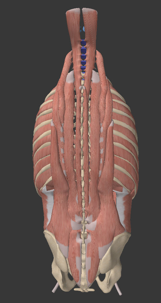
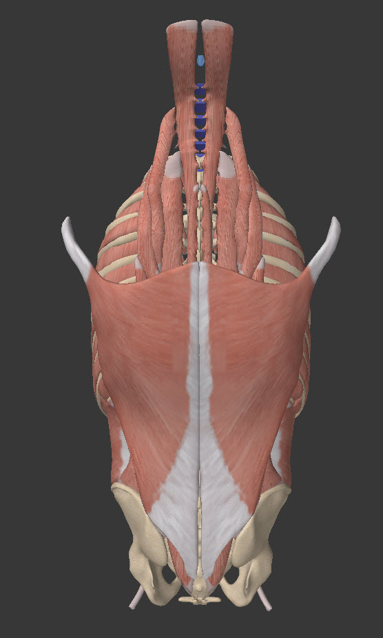
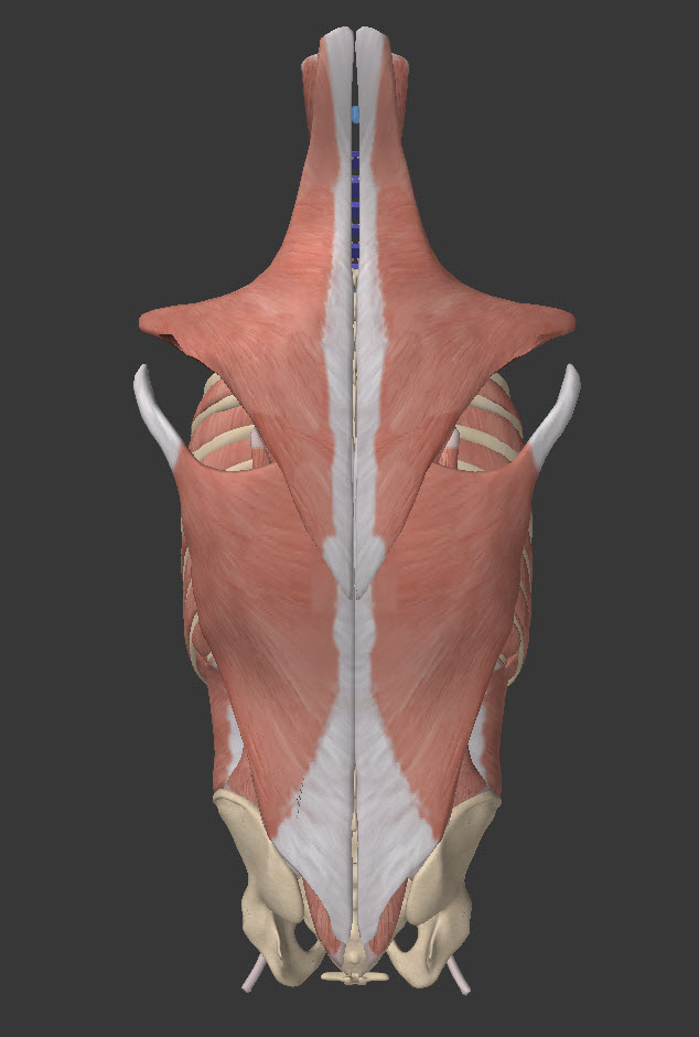
The back extensors are long overlapping columns of muscle |
 |
 |
|
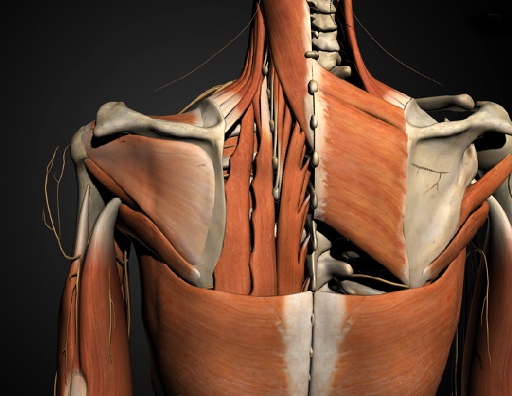
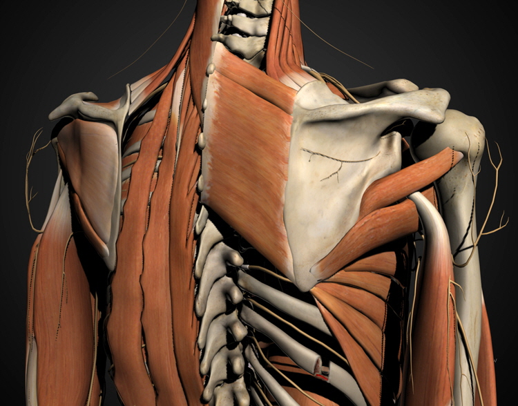
trapezius (upper) & latissimus dorsi (lower) the two great sheets of outer back muscle. The trapezius works the scapulae & clavicles, the lats => upper arms.
The intercostal muscles follow the same plan as the abdominal muscles and can be thought of as an extension of the abdominals broken by islands of bone called 'ribs'. |
|
|
|
The costal cartilage is removed here. In children whose bone is soft, the diaphragm can pull in a line of indentation along the diaphragm edge. In these images the abdominals are seen as smooth and with horizontal direction. This is the middle layer of the three layer (3 ply) abdominal musculature. The outer layer muscle fibers run the way your arms go with your hands in front pockets The deep layer of the three runs the way your arms go with your hands in your back pockets.
Deep to these three is a non-muscular filmy thin but tough layer that lines the muscular cavity - the transversalis fascia. The peritoneum is a wispy thin wet film that lines the true inner surface. It gives its name to the abdominal cavity - the 'peritoneal cavity'.
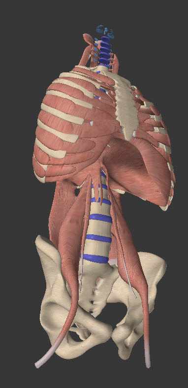
What do you call an intercostal muscle that runs from the lowest true ribs to the top of the pelvis (a rib mass of several fused in developmental sense). That is the quadratus lumborum. Quadratus because it has roughly 4 edges and borum because it is boring - no - wait - lumborum because it is in the lumbar area and Latin people know how to really move this one.
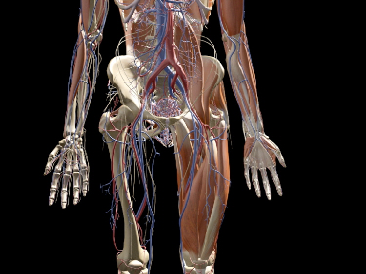
The Iliopsoas muscle (the two act together) is the largest motor of walking and gives walking almost ALL of its energy. Look at the company it keeps. Huge vessels with big contributions at every spinal level. Big red ones in and big blue ones out. The WORK is done here. Heat is given off here. Ooooo. The heat of walking is retained in the body rather than the legs? Yup. The legs work like pendulum parts. The oomph comes from the initial push of the wing (except that it is a pull).
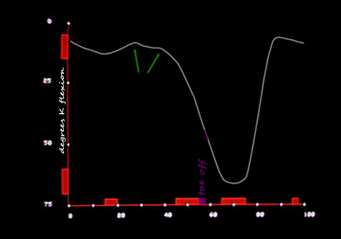
So what is flexing the knee? The iliopsoas. We actually don't SWING the LEG . . . we swing the THIGH. That steep slope down the curve to about two thirds is the thigh swinging and the toe still on the ground! Picture the last part of this curve looking like a saxophone: last 1/3rd of down then steep up then the mouth piece. The sax is a swing instrument. Swing phase as defined by the TOE being off the ground - is saxophone shaped. What is doing that? The inertia of swinging the thigh. The swing of the lower leg is like a towel snap. It is the hand on the other end providing the movement - the hand of the iliopsoas. So officially stance phase is the small curve and most of the down swing of the larger curve (approaching 60 degrees) as the toe pulls away from the dirt. That is simply STUPID ! Why give toes such a big role? The big curve begins (the knee BEGINS to flex) when the THIGH begins to swing. That is happening at the first green arrow as ground reaction and ankle stiffness retards - something that is a bit variable. I originally called this wiggle the curious wobble trying not to be rude to previous authors while calling attention to the fact that commercial devices measuring movement make their data look pretty by smoothing. Bessel & spline smoothing methods which make BEAUTIFUL curves kill this data. What do they kill? They blind us to seeing the resistance to initiation of swing phase caused by the foot and ankle, where easily 60% of Iliopsoas energy can be lost. Nice going guys & gals. MOST gait labs still have this defect (as of Oct 2011).
Polar graphs handle periodic functions better by allowing to better visualize the subharmonics that build the overall big shape. This was studied on mathematical models and on actual patient data and the clear cut removal of data in a very significant range to cause trouble is easily demonstrated this way. Remember that an ellipse is a circle with TWO centers. So an elliptical pattern is not merely two - how about that, there are two of'em - harmonics, but rather one event in which the two harmonics are tightly controlled and keenly paired . Coordinated is a nice word. In polar graph form the big sweep knee motion is an ellipse. The two pendulum components are well matched. Deviations cost energy. Soooo, the muscles in the leg help SHAPE this event, but do not cause it. So it is swing the thigh and flip out the lower leg. Hamstrings stop the lower flip out just as you approach the mouth piece of this curve . Like Michael Finnegan, begin again. And the energy expended and lost in this is kept in the body, warming the innards. And a big player in all this is a back or torso muscle.
|

|
|
 |
||||||||||||||
|
|
||||||||||||||