|
|
|
|
|
||||||||||||||||||||||||||
 |
||||||||||||||||||||||||||
|
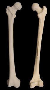
The thigh is the region of the femur. At the proximal (in proximity to the body) is the hip. At the distal end (distant from the body) is the knee. Nobody uses the distinction of 'leg' to mean only a portion of the full leg from hip to toes. Well, maybe one really up tight geezer who also insists on calling cars horseless carriages. |
|
So, the shaft of the femur in resting stance on one foot is not expected to be vertical. Just to get everybody confused, we say that the tibia is in valgus relative to the femur. It's wrong, but we say it anyway - wink wink. Valgus means that as you go distally from the reference joint, the bone deviates away from the body midline. Actually the tibia is dead parallel to that midline and it is the femur approaching it. Relative to the femoral neck, the femur shaft is in varus (approaches the body midline as we go down). That works. Whatever. The back of the femur at the top is deeper relative to the neck than the front.
If the psoas is short and prevents the hip from extending, then the lesser trochanter at that point becomes an unusual fulcrum in that it is not only not the femoral head but it is also posterior. Pressure on the head of trying to extend the leg against this fulcrum presses the femoral hear forward and upward. A psoas contracture can - via leg momentum - act as a fulcrum to have the socket pull a hip prosthesis up and out dislodging it and spinning it forward. That same force in repetitive deformation produces a steeper femoral neck (valgus) and nudges the head forward (anteversion). The sweep of short red arrows indicates that an important bundle of blood vessels sneaks up the back of the neck on its outer surface to enter the femoral head. This is an important inflow of blood to the femoral head. Obstruction or shearing of these vessels will leave the head without blood supply in part or in whole. In the adult the avascular femoral head dies (becomes necrotic) and is called avascular necrosis. However, in a child, as a goodly portion of the head is still cartilage which does not nourish from blood vessels, the necrosis only involves the bone portion. That distinction make the name different - Perthes (for short) disease.
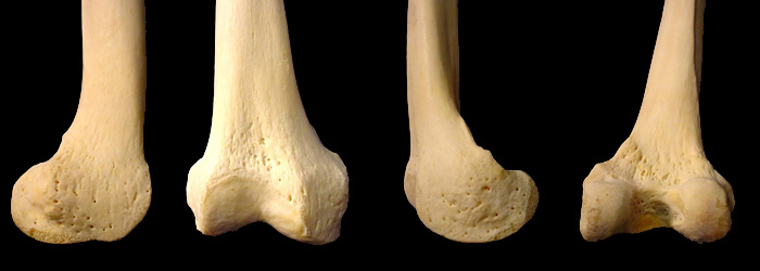
The distal end of the femur is one of the four boney parts of the knee. The others are tibia, patella, and indirectly (by being a ligament handle) the fibula.
Outer (lateral) Front (anterior) Inner (medial) Back (posterior)
The femur is a big bone. A big long bone. It is not dissimilar in architecture overall to the humerus and at its distal joint also to the finger and toe bones. Even the relative valgus of the tibia to the femur is not unlike the "carrying angle" of the ulna to the humerus. One can consider the olecranon portion of the ulna to be the equivalent of the patella.
... mmm ... wonder what's for supper? |
|
|
||||||||||||||||||||
 The femur is a long bone whose axis of movement is well outside of its
substance for most of its length. Indeed, the important relationship of leg anatomy is that a line drawn from the the hip center to the ankle center passes right through knee center. Additionally, with that leg line vertical
the knee and ankle surfaces ought to be horizontal.
The femur is a long bone whose axis of movement is well outside of its
substance for most of its length. Indeed, the important relationship of leg anatomy is that a line drawn from the the hip center to the ankle center passes right through knee center. Additionally, with that leg line vertical
the knee and ankle surfaces ought to be horizontal.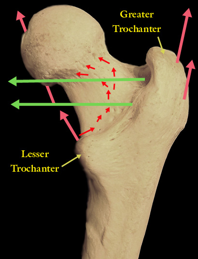 Looking at the back of the right femur
we see the deep ridge that goes from greater to lesser trochanter. The greater is the handle for upward pulling hip abductors (gluteus medius and minimus). The lesser is the handle for the psoas
tendon. Sitting behind the femoral head, the flexion action of the psoas is also an outward rotation force as the lesser trochanter is pulled forward and upward - spinning and raising the femur.
Looking at the back of the right femur
we see the deep ridge that goes from greater to lesser trochanter. The greater is the handle for upward pulling hip abductors (gluteus medius and minimus). The lesser is the handle for the psoas
tendon. Sitting behind the femoral head, the flexion action of the psoas is also an outward rotation force as the lesser trochanter is pulled forward and upward - spinning and raising the femur.