|
BRACHIAL PLEXUS BIRTH INJURY
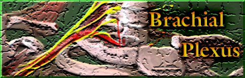
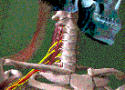 Please review your anatomy. In brief, nerves exit the spinal cord in the lower neck and travel to an area underneath the collar bone, where they coalesce to form the brachial
plexus - almost like a fuse box. It is here that these nerve roots intermingle to emerge as the major nerves which innervate the shoulder, arm, and hand. Those nerve roots are
the fifth, sixth, seventh, eighth cervical nerve roots (cervical = in the neck) and the first thoracic nerve. Occasionally the fourth cervical or the second thoracic root is included.
Please review your anatomy. In brief, nerves exit the spinal cord in the lower neck and travel to an area underneath the collar bone, where they coalesce to form the brachial
plexus - almost like a fuse box. It is here that these nerve roots intermingle to emerge as the major nerves which innervate the shoulder, arm, and hand. Those nerve roots are
the fifth, sixth, seventh, eighth cervical nerve roots (cervical = in the neck) and the first thoracic nerve. Occasionally the fourth cervical or the second thoracic root is included.
 2 .. 3 / 1000
During birth, the baby may become
stuck when the head or the shoulder is
unable to clear the mother's pelvic outlet.
In trying to get the baby delivered before
any serious breathing or circulation
problem occurs, traction on the head or
shoulder may stretch nerve roots.
Traction could even pull one or
more roots out of the spinal cord.
Injuries, such as these, occur roughly 2 to
3 times every 1000 births. They have tra-
ditionally been grouped into three categories:
The first one is the classic Erb's Palsy, a condition resulting from injury during birth which affects the C5, C6, and sometimes C7 nerve roots. This type is the most common type and fortunately has the best prognosis. The children affected have full use of the hand and fingers, but the function of the shoulder, elbow and forearm are affected.
The next category is Total Plexus Palsy, which involves all roots of the brachial plexus. The entire upper extremity is affected.
The last category, Klumpke's paralysis, which occurs in well less than 1% of the cases involves the lower roots (C8, T1). These children have normal use of the shoulder and elbow but weakness and/or paralysis in the fingers and the hand.
Even though we classify these injuries into these three broad categories, that is, into three generalities, the fact is that each patient's injury is typically much too complex to fit totally into this classification system.
To better explain this point, let us digress a bit and go back to some basic concepts. Nerves are basically the electrical wiring in the body. When a nerve gets stretched a variety of changes may occur to its structure; a minor injury may be a temporary "short" to a few nerve fibers within the nerve. ________..____________
If the injury is more severe, there may be complete disruption of the nerve with scarring around the
nerve preventing the healing. ______/` ~~;_________
The nerve fibers around this injury become very disorganized like a tangle of wires and the nerve loses all function.
Another way of thinking about it is, that each injured nerve root or trunk can be like a car that has been in an accident. It can be as mild as a small fender bender where the car will still work and be repaired, they can be so severe that the car(or nerve) will never work, or anywhere in between those two.
 The brachial plexus injury may be as minor as a temporary paralysis
that completely recovers or it may be so severe that the entire limb is permanently paralyzed. It is difficult to tell by the diagnostic tests available today, the exact nature and extent of the injury and its
potential to heal and recover. That is why initial management is a wait and see. But wait how long? And, see what?
The brachial plexus injury may be as minor as a temporary paralysis
that completely recovers or it may be so severe that the entire limb is permanently paralyzed. It is difficult to tell by the diagnostic tests available today, the exact nature and extent of the injury and its
potential to heal and recover. That is why initial management is a wait and see. But wait how long? And, see what?
Unfortunately for prognostication (predicting what will be), the degree and mix of injury, and the magnitude and rate of recovery vary from patient to patient. We, therefore, believe that each patient should be individually evaluated and a regimen of treatment prescribed which addresses each child's individual deficits.
There are certain nuances to the natural history of this condition that if ignored will have significant impact on the final result.
Although roughly 85% of the patients regain "full" neurologic recovery by three or more years, the delay in normal function and the presence of any muscle imbalance across any joint has significant impact on the growing skeleton.
A child may be left with permanent musculoskeletal abnormalities. I believe that every effort must be made to maintain full motion of all joints, primarily through a supervised therapy program and when necessary assisted with surgical intervention.
Timing is important. It is important to understand that there are windows of opportunity for all treatment interventions to positively impact on the final result. Therefore, these patients ought to be followed at regular intervals to monitor for neurologic based muscle recovery on the upside and development of joint contractures on the downside.
With knowledge, one can avoid pitfalls and optimize a patient's outcome within the range of possibility.
When an infant in the newborn nursery is noted to lack movement of the upper extremity, a physical exam determines the presence or absence of non-neurologic injuries. Birth trauma may fracture the clavicle, humerus, ribs, or even the femur. A limb fracture will behave as pseudoparalysis, that is, the baby won't move the injured limb. A fracture alone may make the limb seem paralysed as the baby's protective mechanism shuts down usage.
Again, as in autos, a headlight broken does not exclude a wiring loss to that same headlight. Bulb replacement might not result in luminance.
Brachial plexus injury may occur along with fracture. In the presence of a fracture the infant is protected (by, say, pinning the extremity to the torso by sling and swathe) for about 10 days before enough healing has occurred to begin therapy. Though rare, acute shoulder or elbow dislocation can occur at birth. This needs to recognized and treated as well.
When brachial plexus injury has occurred without an associated fracture or dislocation, traditionally, the affected arm has been, in similar manner, pinned empirically (the idea being to protect the recently injured plexus). There is no data supporting that. We believe that immediate gentle restricted motion should be instituted, and no pinning or immobilization is routinely done. Stiffness is the problem. That's what we seek to preempt.
In the natural history of brachial plexus birth injuries,
recovery of the injured plexus and nerves may occur
over the first 6 years of life.
During this time
the child may completely recover
or
the clinical progress may plateau.
Our goal is to maintain full motion of all joints, primarily through an ongoing intense therapy program. If neurologic recovery falters or plateaus, there are certain specific indications for exploration and nerve grafting that have emerged.
Amongst physicians treating these injuries, there is great disagreement about the timing and indications for such surgery. This is partly the result of the history of brachial plexus injuries.
In 1927, Sever published a paper evaluating 1100 consecutive
brachial plexus birth injuries. He saw no difference in outcome
between the operative and non-operative patients. As a result,
treatment of this condition remained largely conservative for
over 50 years.
The entire management approach evolved into "wait and see".
While these children generally adapted and functioned in society,
changes in attitudes toward disabilities as well as the advances in
microsurgical techniques, intraoperative electrophysiologic moni-
toring, led to reexamination of the approach, with renewed interest
in intervening in these sometimes-debilitating injuries. So, the treat-
ment of brachial plexus birth injuries is really still in its infancy.
Medical science is still trying to work out the optimum management
of this condition.
In our center, we believe that:
Babies with Total
Plexus Palsy
showing no recovery by 3 months, ought to have their brachial plexus surgically explored and, if suitable for it, nerve grafted.
Children with Erb's Palsy
without strong elbow flexion or shoulder elevation by 6 months, should have surgical exploration and nerve grafts performed. Alternatively, depending on what is found, muscles can be rearranged (muscle
transfers) to offset the effects of anticipated permanent losses.
![]() Due to the nearly universal involvement of the C5 and C6 nerve roots, children
with brachial plexus birth injuries usually have some impairment of the function of the shoulder and elbow. The purpose of the shoulder is to position the hand in
space. Recent studies have demonstrated the profound effect of limited shoulder motion has on the function and strength of hand.
Due to the nearly universal involvement of the C5 and C6 nerve roots, children
with brachial plexus birth injuries usually have some impairment of the function of the shoulder and elbow. The purpose of the shoulder is to position the hand in
space. Recent studies have demonstrated the profound effect of limited shoulder motion has on the function and strength of hand.
Joints require full motion to develop normal anatomic shape and function. The growth and development of the joint surface is an ongoing process that involves active growth on each side of the joint. Depending on the nature and extent of recovery, muscle weakness or lopsided muscular action around any joint in the arm may develop. Either of these may cause joint deformity and loss of function.
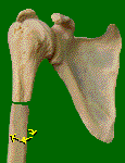
The Shoulder
The shoulder joint has the most motion of any joint in the body. 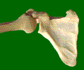 Elevation of the shoulder is a
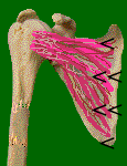
complex motion involving a coordinated combination of two joints, glenohumeral and scapulothoracic motion. The gleno-humeral joint is the articulation between the arm
bone and the shoulder blade.
Elevation of the shoulder is a
complex motion involving a coordinated combination of two joints, glenohumeral and scapulothoracic motion. The gleno-humeral joint is the articulation between the arm
bone and the shoulder blade.
Click this image to play movie:
 |
The scapulo-thoracic joint consists of the muscle coated underside of the shoulder blade riding upon
along the outer surface of the rib cage. Researchers have attempted to determine the minimal requisites of raising the shoulder in the air and the![]() role and relative importance of specific muscles. One essential
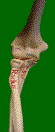
condition for elevation is agreed upon: active external rotation of the shoulder, or being able to
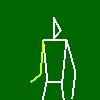
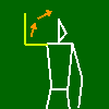
swing the arm out, is an essential requirement for normal shoulder elevation and the ability to bring the hand to the face. Without outward rotation, getting the hand to the face requires the clarion position (fig. on right).
role and relative importance of specific muscles. One essential
condition for elevation is agreed upon: active external rotation of the shoulder, or being able to
swing the arm out, is an essential requirement for normal shoulder elevation and the ability to bring the hand to the face. Without outward rotation, getting the hand to the face requires the clarion position (fig. on right).
Click on this image to see clarion movie:
 |
If internal rotation contracture of the shoulder develops and persists, pathologic changes in the glenohumeral joint ensue with possible subluxation or dislocation, resulting in restricted motion and function.
Synonyms : ER, external rotation, outward rotation, lateral rotation
IR, internal rotation, inward rotation, medial rotation
Passive motion must be maintained in order to ensure the development of a congruent glenohumeral joint. [congruent = both sides of the joint have shapes which match each other through the functional range of movement. For example, a hip which has egg shaped parts, is only congruent for movement in one plane, typically flexion, but incongruent in motion in other directions such as abduction or rotation on the long axis. Incongruence is suggested by lack of concentric centers. That is, the center of the ball ought to coincide with the center of contour of the socket.]
With the progression of ossification of the humeral head and glenoid, the shoulder joint takes on its permanent shape at about 3 years of age. Therefore, if our therapy is unable to maintain external rotation and/or a concentrically located ball in the socket, then surgical intervention is necessary before age 3 to establish joint congruence and thus, avoid a mechanical permanent loss of motion.
It is for these reasons that we initiate a carefully supervised physical therapy program utilizing specially
talented therapists knowledgeable in special needs of such children. They educate parents and care givers in the techniques proper for joint motion preservation.
To maintain external rotation (considering description in a theoretical
standing posture, standing or not), the therapist or caregiver performs this exercise with the upper arm held against the side of the torso. Outward
rotation of the arm swings the forearm forward to pointing straight ahead. Abducting the shoulder (moving the elbow away from the body) to 90 degrees would have the forearm still parallel with the
floor. That does not 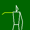
 get the maximum stretch out of the anterior shoulder capsule nor the muscles that
rotate the arm inward toward the body.
get the maximum stretch out of the anterior shoulder capsule nor the muscles that
rotate the arm inward toward the body.
Additional external rotation raises the forearm to point at the ceiling Furthermore, all motions of the upper extremity are attended to, with
special care taken to isolate glenohumeral motion from scapulothoracic motion. It is important to maintain a continuous physical therapy program at home and with a
therapist; interruption of this program will lead to rapid loss of function and contracture.
The experience and creativity of the therapist is critical when attempting to cajole a young patient in the non-compliant phase of development to perform therapeutic exercises designed to maintain motion and gain strength. Play activities are designed to incorporate the desired motion and exercises. In cases of complete plexus involvement where wrist drop or finger flexion contractures are a concern, a cock-up splint is used.
We also incorporate electrical stimulation into our therapy both with the therapist and at home, to
minimize permanent muscle atrophy. The use of electrical stimulation in these patients is a new technique that sends small amounts of current into the muscle. While there is no definitive proof of its
efficacy, the thinking is that it stimulates the muscle to prevent it from completely withering away and it may even stimulate nerve growth.
In the absence of shoulder dislocation, if external rotation cannot be achieved through the physical therapy program, then surgical release through a subscapularis slide (an advancement of the muscle that blankets the undersurface of the scapula reaching laterally to attaches to the front of the humerus) is performed usually at the age of 18 to 24 months. The child attains, with that, immediate improvement of both function and cosmesis.
If over the course of the ensuing year the contracture begins to recur, a modified L'Episcopo procedure is performed, where the latissimus and teres major insertions are transferred around to the back of the humerus into the infraspinatus tendon. These muscles are powerful internal rotators of the
shoulder that join together in what is called a conjoint tendon which attaches into the front of the arm bone or humerus. This transfer is possible because the shoulder has
additional powerful muscles which perform similar movements akin to those of the transferred muscles. This transfer will permanently balance the muscles about the
shoulder thus preventing recurrence of the contracture and improving elevation of the shoulder.
 When permanent bony incongruence has developed in the glenohumeral joint, in
those patients who are seen late, an external rotation osteotomy is performed to optimize the position of the arc of motion and improve function and appearance (Figure on left).
When permanent bony incongruence has developed in the glenohumeral joint, in
those patients who are seen late, an external rotation osteotomy is performed to optimize the position of the arc of motion and improve function and appearance (Figure on left).
When minimal muscle recovery occurs and there is little or no ability to elevate the shoulder. Some salvage procedures have been proposed including shoulder fusion and trapezius transfer. In our center, we believe that shoulder fusion is the last resort. The trapezial transfer leaves the patient with limited abduction, a webbed neck and a poor cosmetic appearance. Thus, in the face of profound abduction weakness. I prefer to perform a complete transfer of the latissimus dorsi, which in addition to gaining abduction power also gives some bulk to the shoulder area and improves the cosmetic appearance of the shoulder.
The Elbow
 In many children with Erb's palsy, a mild paradoxical elbow flexion contracture often develops.
To date there is no definitive explanation of when and why this occurs. I believe it occurs when there is some weakness of both the elbow flexors and extensors, and a contracture
develops to give some mechanical advantage to the flexors, which are more essential for function. In any event, it is also difficult to undo. I have used a spring-loaded extension splint
for evening or night use that may partially decrease the flexion contracture.
In many children with Erb's palsy, a mild paradoxical elbow flexion contracture often develops.
To date there is no definitive explanation of when and why this occurs. I believe it occurs when there is some weakness of both the elbow flexors and extensors, and a contracture
develops to give some mechanical advantage to the flexors, which are more essential for function. In any event, it is also difficult to undo. I have used a spring-loaded extension splint
for evening or night use that may partially decrease the flexion contracture.
When biceps power lacks the ability to flex the elbow against gravity, a significant functional impairment exists; the patient cannot bring the hand to the mouth for feeding, buttoning a shirt, and combing the hair. Muscle transfers when neurologic recovery has plateaued, will give the patient better ability to use the hand. If minor weakness is present, a Steindler flexorplasty is performed [where the origin of flexor wad of the forearm is moved proximally and centrally to assist in elbow flexion]. If more profound elbow weakness is present, several procedures have been in existence to gain function. My preference is to do a latissimus dorsi transfer [ a muscle from the back is swung pivoting on its upper tendon into the arm].
![]() When significant imbalance of the muscles that rotate the forearm are present early in recovery, a pronation (palm down) or supination (palm up) contracture may develop
which limits function and sometimes gives an unsightly cosmetic appearance. Even if the opposing muscles recover sufficient power, it, alone, is not capable of reversing a fixed contracture. It is important to release these contractures BEFORE fixed bony deformity occurs in the radius and ulna (the 2 bones of the forearm). When these subtle changes occur, the rotation of the forearm will be
permanently restricted. Therefore, a window of opportunity exists in the first few years to release forearm contractures and regain function of the forearm.
When significant imbalance of the muscles that rotate the forearm are present early in recovery, a pronation (palm down) or supination (palm up) contracture may develop
which limits function and sometimes gives an unsightly cosmetic appearance. Even if the opposing muscles recover sufficient power, it, alone, is not capable of reversing a fixed contracture. It is important to release these contractures BEFORE fixed bony deformity occurs in the radius and ulna (the 2 bones of the forearm). When these subtle changes occur, the rotation of the forearm will be
permanently restricted. Therefore, a window of opportunity exists in the first few years to release forearm contractures and regain function of the forearm.
In a classic Erb's palsy, involvement of the C5 root leads to weakness of the biceps brachii. In
addition to being an elbow flexor, the biceps muscle is the most powerful supinator of the forearm, which rotates the palm up. With significant weakness of the biceps, a pronation contracture may develop which limits function in hygiene, toileting, and sports activities. If the biceps recovers significant power we have
demonstrated lasting improvement in function by releasing the pronation contracture in the forearm (ref = Liggio et al).
brachii. In
addition to being an elbow flexor, the biceps muscle is the most powerful supinator of the forearm, which rotates the palm up. With significant weakness of the biceps, a pronation contracture may develop which limits function in hygiene, toileting, and sports activities. If the biceps recovers significant power we have
demonstrated lasting improvement in function by releasing the pronation contracture in the forearm (ref = Liggio et al).
Conversely, if the biceps recovers enough in a patient with complete plexus injury, the muscle imbalance may cause a supination contracture of the forearm, which is cosmetically unsightly and impairs use of a keyboard, which in today's computer world is quite problematic. If that occurs and is to be treated before bony changes occur to the radius and ulna, an interosseous membrane release can be performed.
If there is no significant recovery of the pronators of the forearm, which rotate the palm down, a rerouting of the biceps insertion (to reverse the wrap on the radius) will assist in pronating the forearm to neutral pronation (Zancolli procedure).
If a treatment is sought late - with contracture and permanent bony changes in the radius and ulna - then rotational osteotomy of these bones to position the forearm in neutral is indicated.
In patients with Total Plexus Palsy or Klumpke's paralysis, the hand and fingers are affected. The nature of the involvement is highly variable and can include a number of deficits, which require individual assessment for, if necessary, tendon transfers or releases. There are myriad possibilities, too many to generalize. Each child must be individually evaluated and managed with a combination of therapy, splinting and occasionally surgery.

There are facial findings that may be found at birth in cases of brachial plexus injury. Sagging eyelid and pupil size mismatch suggest an additional injury deeper than the plexus itself. Also note that some  embryologic - that is genetic type - disorders of
formation can mimic brachial plexus injury. These are related to embryologic gill formation.
embryologic - that is genetic type - disorders of
formation can mimic brachial plexus injury. These are related to embryologic gill formation.

summary, the management of the child with brachial plexus injury is an ongoing task that requires individualization of treatment and is impacted by the severity of injury,
the rate and degree of recovery of the muscles, and the age when the child appears in our program for initiation of treatment. Optimization of passive motion while neurologic recovery is occurring is
essential for the normal development of joints; this is primarily achieved through ongoing therapy. Surgical intervention is necessary during this period if neurologic recovery falters, if shoulder
subluxation or dislocation occurs, or contracture becomes fixed. Once recovery has plateaued, muscle releases and/or transfers may better balance the joints and improve function and appearance.
