
Basic Anatomy
Between the central nervous system (brain & spinal cord) and the end organs innervated (muscles, glands, sensory organs, etc.), organization is quite tight. It is amazingly regular and orderly.
It is the getting there, passing through and around anatomy, where neurologic anatomical variation is most seen. Tiny hair-like nerve rootlets, each carrying small clusters of neurons come off the spinal cord like the teeth of fine tooth comb. To get where they are going as organs spring up embryologically, these regular they are grouped into bunches - OK, everybody, pick a bus, we'll meet under the umbrella when we get there - and complete their journey.
A single muscle may well have three different nerves that act as conduits of its innervation. Yet the central organization does not reflect the circuitous route that one or another nerve might take. How long have you been here? Our bus had to take a detour.
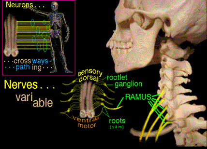 So, think of nerves as roadways and innervation as commercial traffic, free to take whatever route gets there. Of course, some routes can be expected with regularity as they are better suited.
In the low neck & shoulder area, this New Jersey clover leaf traffic pattern is at its ultimate in what we call the brachial plexus. Brachial means 'of the arm' in Latin. Plexus means 'constructed of folds or windings' as
related to 'complex'. So when neurons go from here to there, anatomists in a fury of synonyms try to give each individual grouping or branch an impossible name to remember. Job security.
10 Rootlets: The neurons come off the spinal cord in absolutely regular tiny hair like groups called rootlets. Those coming from the back side of the spinal cord are sensory, those from the belly side of the spinal cord are motor rootlets (5 each).
5 Roots: The rootlets converge into groups even before getting through the outer spinal cord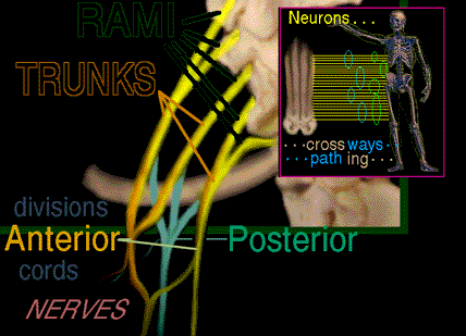 wrapper (meninges) and form sensory (or dorsal =
backside) roots and motor (ventral = belly side) roots. Because cell bodies tend to be at the feather end of the neuron arrows, sensory cell bodies tend to be outside the cord and motor cell bodies inside the
central structure. Thus sensory ganglia (clusters of cells which serve the neurons) are seen with the dorsal sensory roots and not with the motor (motor has them too, but they are upstream inside the central
system). Some folks also call these roots "rami". Others reserve the root term for other regions where this mish mosh isn't going on. OK, so 5 rami, too. We'll pick it up as rami.
5 Rami: (pleural of ramus = branch in Latin, ignoramus = one who can't or won't follow
complexity)
Going my way? Even though sensory neurons point centrally and motor neurons point peripherally, as with roads, traffic can be two way. Where the dorsal and ventral roots (sensory
and motor) share a single conduit has been called [at least nearest the spine] rami. After the roots give off some local traffic, each ramus has sensory and motor lanes. But rami are really
two way access lanes in and out of the vertebral entry ramps into the spinal canal. A ramus, being just an easy passage from the spinal canal, has little specialized organization. That is,
other than the mix of sensory and motor, there is little destination sorting. Rami reflect the orderly spinal pattern of emergence of rootlets and - more or less - group them for passage out and that's it.
There are about 5% of people who are prefixed or postfixed. That means that some or all of the here to there traffic is exiting the spinal canal one level higher or one level lower. That does
NOT change function at all as the start and finish are not what's different. What is different is just the route taken to get there. What makes this important is that the - say C5 - innervation
may well be coming out with the C6 rami in SOME people. That's why doctors want all those pesky tests for BOTH neurologic identification AND anatomic localization.
3 Trunks: Pack your trunks, a neuron's gotta do what a neuron's gotta do. Rami have to pass the vertebral port holes called foramina. So they're similar sized. But once out of the tunnels, the roadways reflect business. Trunks are the first sorting of neurologic roads based on port of call. The first two (C5 & C6) and the last two (C8 & T1) rami join. The corresponding segment of the middle ramus, although it doesn't join anybody is - so it won't be sad - also called a trunk.
Ramus Trunk
C5
\___________
/
C6
C7___________
C8
\__________
/
T1
Trunks are heading to the arm, but there is front of arm and back of arm. So...
6 Divisions: Each of the three trunks splint in two. One each to the front and one toward the back - anterior and posterior divisions.
3 Cords: Somebody yells, "Hey I know a short cut through the pass." and the three posterior divisions join into one cord. The upper cord convinces the middle division - so far aloof - to join into a lateral cord. The lowermost cord goes it alone renamed as the medial cord.
You following this???? There's more. Sorry.
5 Nerves: Nerves? Hooray. Finally! Anyway the anterior 2 cords continue on but send off connections to form a middle - and very important - third part.
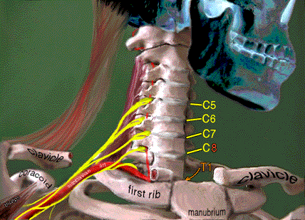 Relative to other structures we see the rami between the bone of the vertebrae
and the edge of the muscle on which the rami sit - the medial scalene muscle (scalenius medius).
The trunks are just past that, ending just
before the nerve components embrace the subclavian artery. That artery comes off the aorta and arches over the first rib to get to the arm (that's the pulse you feel in the arm pit). For visibility, the
clavicle (collar bone) is cut out here.
The divisions begin right at the embrace
of the artery (doesn't embracing always cause division?).
The trunks sit deep to the collar bone.
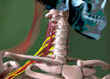 So as the subclavian (means beneath clavicle) artery passes out of the neck
<===== between the
scalenius anterior and the
scalenius medius,
the trunks form divisions and these find accord at the acromion and come together as cords.
The reflected head of the biceps (bi = 2, biceps because it has two heads , one attached to the coracoid) is seen right. Right there, the coracoid points to the top of a letter M. The outermost leg of the M is the musculocutaneous nerve. The middle leg is the median nerve. The innermost leg is the ulnar nerve. Some cutaneous nerves also come off right about here as do some pectoral nerve branches further up. But we are looking at the big stuff here.
 Ptosis of the eye (sagging of the eye lid) is an important finding. The tiny rootlets which exit the spinal cord have some portions
diverted to travel upward within the spinal canal and not exiting between the vertebrae. Those upward traveling rootlets group into a nerve that enters the skull alongside the spinal cord. These carry
control signals for the pupil of the eye and for the eye lid raising muscle. To damage these, which are not in a direct line of tension with the brachial plexus outside the spinal canal, means that injury
was transmitted deep within the spinal canal. That injury was avulsion of nerve root and rootlets from the spinal cord itself. That is ominous indeed. MRI of the spinal cord will probably
show spinal cord damage in addition to plexus damage in such an instance. But note, there are lesser unrelated things which can also make an eyelid sag. The first long
branch to come off the brachial plexus is the long thoracic nerve (going to the serratus anterior). If that muscle is paralyzed, then the plexus injury is very likely close to the spine (or within it).
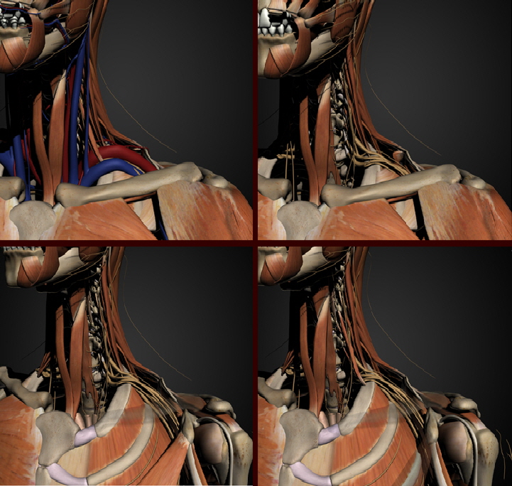 |
Removing things that block our view we see that the brachial plexus roots C5, C6, C7, & C8 do not come from the thorax, though C8 teeters on it and in cases of extra ribs does do so. T1 root has to travel upward to get over the first rib.
With the clavicle (collar bone) and pectoralis minor ghosted (and pectoralis major removed , we see the route of the brachial plexus into the upper arm. En route, portions of the roots swap content with each other forming the intersecting ramps of this highway we call the brachial plexus.
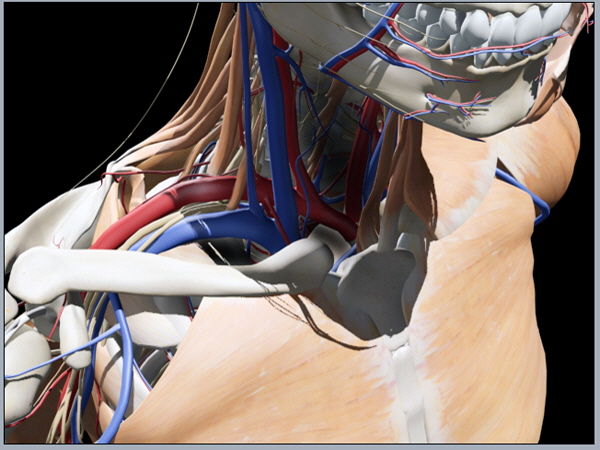
Here's another view with many things removed to show the boundaries of the path of the brachial plexus.
Now imagine being thrown from a motorcycle and landing such that the head goes to the far side while the shoulder is driven 'downward' (caudal, as down may be up in this instance).
Dr. Nuzzo horns in here...
Conclusion: Watch out for cars driven by deaf old geezers who have tunnel vision and also seem to have their sole purpose in life to destroy people on bicycles - motor or otherwise.
OK. OK. That was naughty to say. Totally true but - naughty. The alternative is to tell bikers to STOP WEARING BLACK! It does not stand out to those with poorly intact vision and totally intact accelerators.
