CPT, Fibrous Dysplasia, Unicameral Bone Cysts . . .
Are there linkages? Some very peculiar cases to feed curiosity are here.
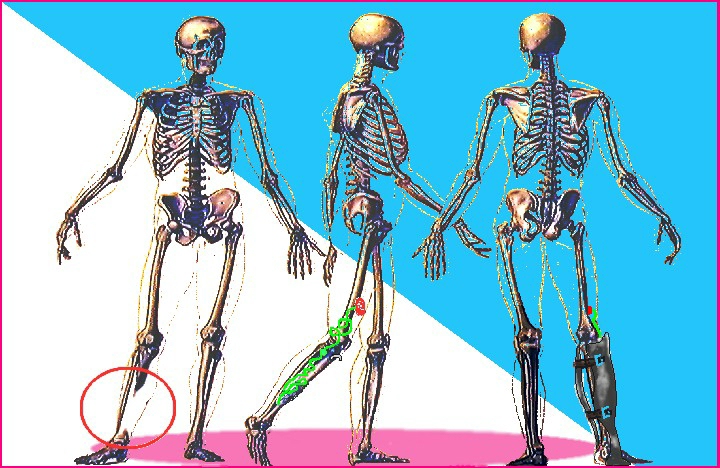
Click on above image to see PDF
Case reports are the bad boys of medicine. They send you on wild goose chases (& publications hate them for that reason). But they can provide rare glimpses of deep quirky mechanisms that rarely otherwise show themselves. They can be a hint as to where to look for answers if not giving answers themselves. In any event they can be more interesting than watching daytime television.
Following are a few more illustrations for those of you more curious about some of the above details. Followed so many years there are too many to be anything but sampled. This sample has a bias as to not seek out the oddball 'winner' images.
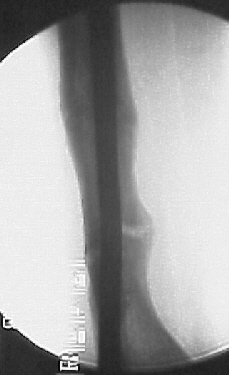
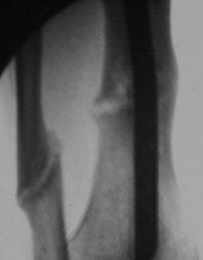
The lesser case of CPT - not disappearing bone but rather multifocal (more images):
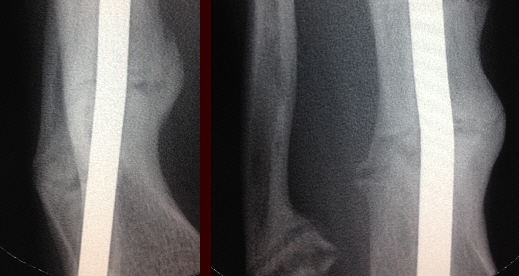
Above, left, we see that the big CPT is well healed and remodeled. You might not even suspect it was the main problem. So much so that thes others were left alone. The lesser defect became painful as the patient ambulated busy NYC streets for school. The lesser defect scraped out on one side only and treated with calcitonin by catheter infusion has healed well only where so treated. The other side had not healed. This is despite the same weight bearing and rod as the main defect. The image on right is magnification view rotated to best see lucency. One of the fibula defects is seen as well (so defect is not JUST tibia.)
At surgery we find the lucency to not be a space but a poorly calcified zone of cartilagenous looking and fibrous tissue. Scraped out and treated with an onQ source of calcitonin (alternate days) it healed as well (not the fibula which was not so treated).
 |
Fibrous dysplasia??
et tu?
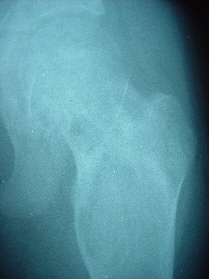 |
This femur broke repeatedly under minimal weight (even with crutches). The bone was too soft to even attempt internal fixation (tibial traction was required for the neck & intertrochanteric fractures). A hypodermic needle was able to inject solution directly into this bone as if it were an orange.
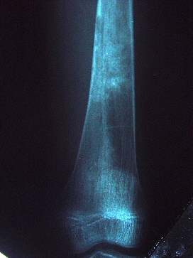
At normal x-ray exposure (perfect for other leg) we see this.
From the femoral head to the knee, there is no area spared. That cinches diagnosis of fibrous dysplasia. But some bone areas look - mmmm - dissolved? Not just not formed. Removed? Invaded & replaced? Who removes and replaces bone in the cast that plays the role of bone?
[See the document at the top pf this page]
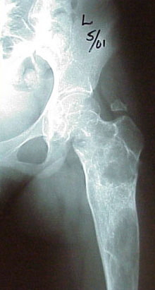 Some spillage of the 'caulking' that was used to fill this bone after it
was hollowed out is seen floating above the greater trochanter. A long tube was used to fill this cannoli-like with calcitonin soaked bone sand (bone had been ground to fine sand and let dry with calcitonin. Then
calcitonin used to suspend the sand at time of filling of reamed center.
A pesky area at the inferior head neck junction defied packing directly (thoracoscope from lateral). Dr. Rosenthal (Harvard Radiology) injected this nast bit for us and it too filled in.

This bone is what followed the calcitonin filling treatment (similar x-ray penetration).
Resumption of controlled ambulation was followed by normal weight bearing and eventually full activity short of spitting into the eye of fate (no parachute anything etc).
The clinical response to calcitonin,a reversal in the direction things were going, makes one more than curious as to the role of osteoclasts or at least to an osteoclast catabolic mechanism in cells to which it ought not be known.
The effect of calcitonin on UBC's turned out to be even scary.[see PDF at top]